グリシン酸マグネシウム 図
6 査読済み研究からの図表
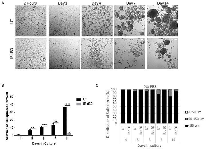
Sphere-forming efficiency of parotid-derived cells decreases significantly following a single 5 Gy radiation dose, quantified at day 30 post-irradiation.
Administration of growth factors promotes salisphere formation from irradiated parotid salivary glands.
Proliferation rates of salisphere cultures from untreated and irradiated parotid glands are compared under serum-free conditions, showing similar growth kinetics despite radiation damage.
Administration of growth factors promotes salisphere formation from irradiated parotid salivary glands.
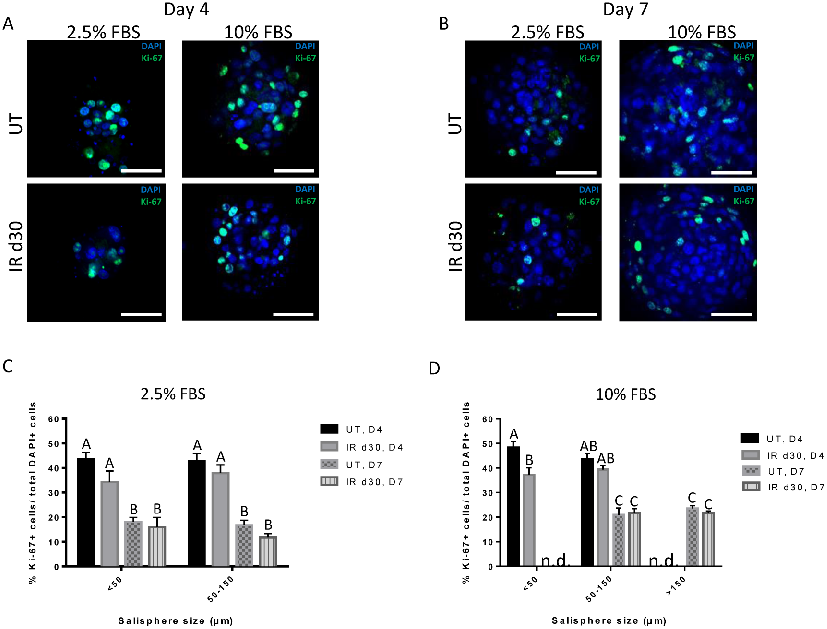
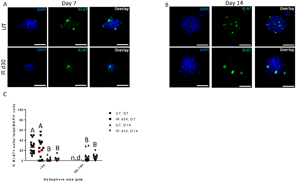
Proliferation assays confirm comparable growth rates between untreated and irradiated salisphere cultures supplemented with FBS, indicating that reduced sphere formation is not due to proliferation deficits.
Administration of growth factors promotes salisphere formation from irradiated parotid salivary glands.
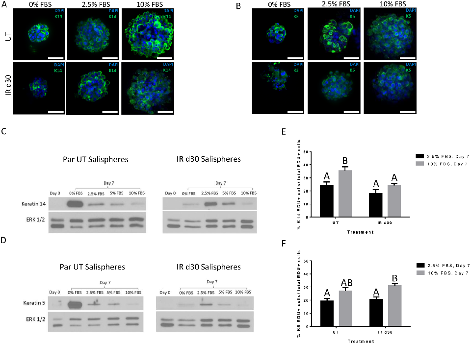
Expression of salivary stem and progenitor cell markers in salisphere cultures demonstrates that both untreated and irradiated cells maintain their progenitor phenotype.
Administration of growth factors promotes salisphere formation from irradiated parotid salivary glands.
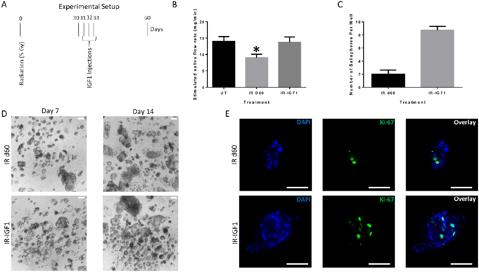
Post-radiation IGF-1 treatment significantly enhances sphere-forming efficiency of irradiated parotid cells, suggesting growth factor administration as a strategy to restore salivary function.
Administration of growth factors promotes salisphere formation from irradiated parotid salivary glands.
Skin hydration measurements in human subjects receiving oral eggshell membrane supplementation are tracked over the study period.
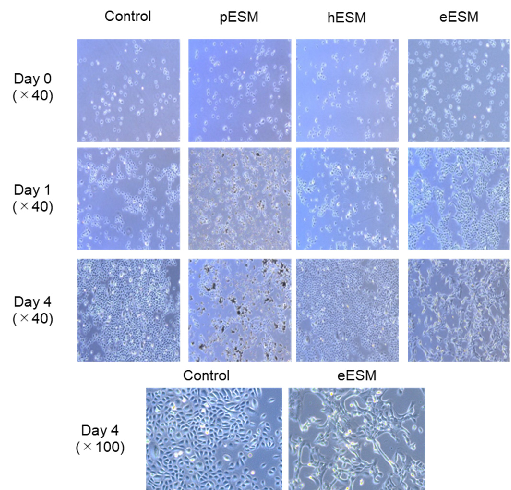
Effects of Eggshell Membrane on Keratinocyte Differentiation and Skin Aging In Vitro …