अनुसंधान प्रक्रिया
14 सहकर्मी-समीक्षित शोध से आंकड़े
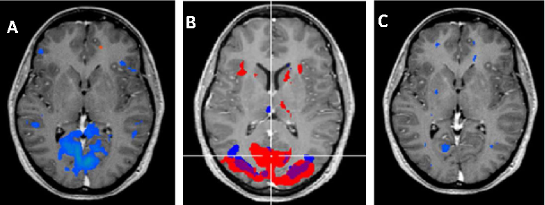
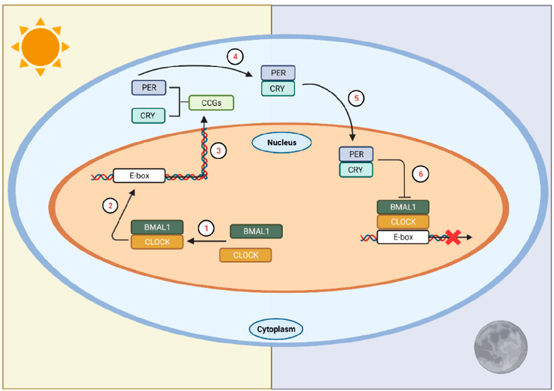
Circadian rhythm regulation involves a complex interplay between the central clock in the suprachiasmatic nuclei and peripheral oscillators throughout the body. Melatonin serves as the primary hormonal signal conveying darkness information to these systems.
New perspectives on the role of melatonin in human sleep, circadian rhythms …
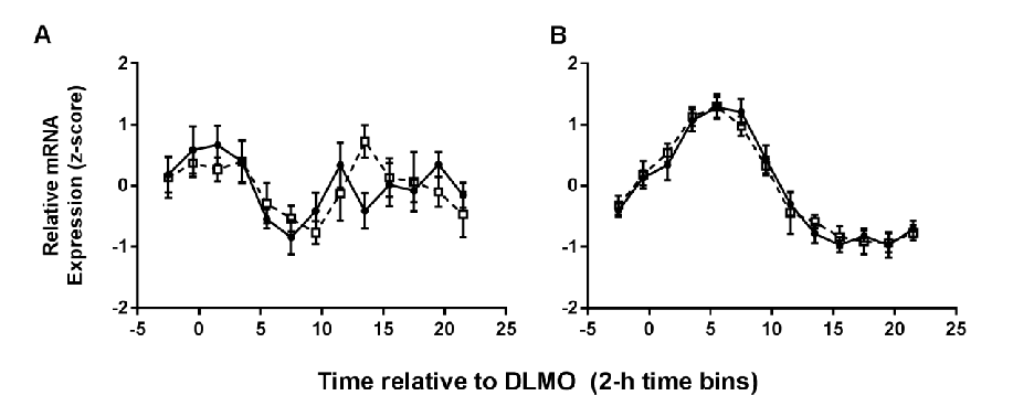
Summary model of how meal timing acts as a zeitgeber for peripheral clocks independent of the central suprachiasmatic nucleus pacemaker.
Meal Timing Regulates the Human Circadian System.
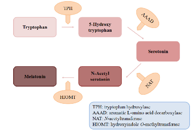
Biosynthetic pathway of melatonin from tryptophan is displayed, showing the sequential enzymatic steps through serotonin N-acetyltransferase and hydroxyindole-O-methyltransferase.
Dietary Sources and Bioactivities of Melatonin.
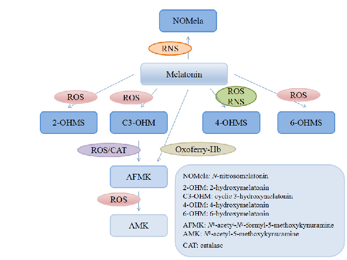
Melatonin and its metabolites including 6-hydroxymelatonin, AFMK, and AMK are structurally depicted, illustrating the biotransformation cascade.
Dietary Sources and Bioactivities of Melatonin.
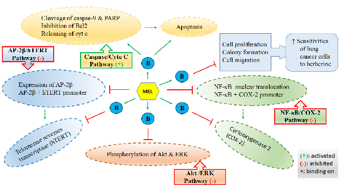
Mechanisms by which melatonin enhances lung cancer cell sensitivity to berberine are diagrammed, showing synergistic effects on apoptotic and autophagic pathways.
Dietary Sources and Bioactivities of Melatonin.
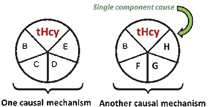
A causal model illustrates how elevated plasma homocysteine may contribute to dementia through multiple pathways, interacting with other risk factors such as age, hypercholesterolemia, and genetic predisposition. No single factor is sufficient alone; rather, combinations of component causes drive disease.
Homocysteine and Dementia: An International Consensus Statement.
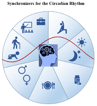
Factors influencing the rest-activity circadian rhythm and the sleep-wake cycle are mapped, encompassing light, melatonin, physical activity, and social timing cues.
Biological Rhythm and Chronotype: New Perspectives in Health.
Metabolic consequences of circadian disruption include altered glucose tolerance, lipid metabolism, and energy balance. This figure summarizes the pathophysiological links between circadian misalignment and metabolic disease risk.
Circadian Rhythms Disrupted by Light at Night and Mistimed Food Intake Alter …
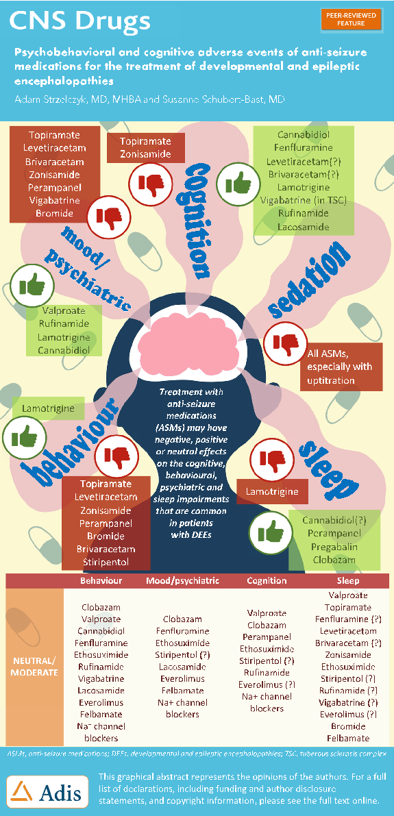
Developmental and epileptic encephalopathies involve severe drug-resistant epilepsy with significant neurodevelopmental comorbidities. This figure summarizes the psychobehavioural and cognitive adverse events associated with anti-seizure medications used to treat these rare childhood-onset syndromes.
Psychobehavioural and Cognitive Adverse Events of Anti-Seizure Medications for the Treatment of …
![Figure 1. One-carbon metabolism. Abbreviations: PLP, plasma pyridoxal phosphate; MTHFR, methylenetetrahydrofolate reductase; FAD, flavin adenine dinucleotide; FMN, flavin mononucleotide. Adapted from [16].](https://pdfs.citedhealth.com/figures/27854316/97.png)
One-carbon metabolism pathways involving folate, vitamin B12, and vitamin B6 are mapped, showing key enzymatic reactions catalyzed by MTHFR and the roles of FAD and FMN as cofactors in homocysteine recycling.
Causes, Consequences and Public Health Implications of Low B-Vitamin Status in Ageing.
![Figure 1. The iRISA syndrome [76] in addiction is centrally controlled by dopamine in the brain, while asynchronization, presumed to be linked to cue sensitivity in digital addiction [50–54], is centrally controlled by serotonin. A deficit in both neurotra](https://pdfs.citedhealth.com/figures/35682491/101.png)
The impaired Response Inhibition and Salience Attribution (iRISA) syndrome model is illustrated, showing how dopamine-mediated reward pathways in the brain drive cue sensitivity in digital addiction and contribute to sleep disruption.
Digital Addiction and Sleep.
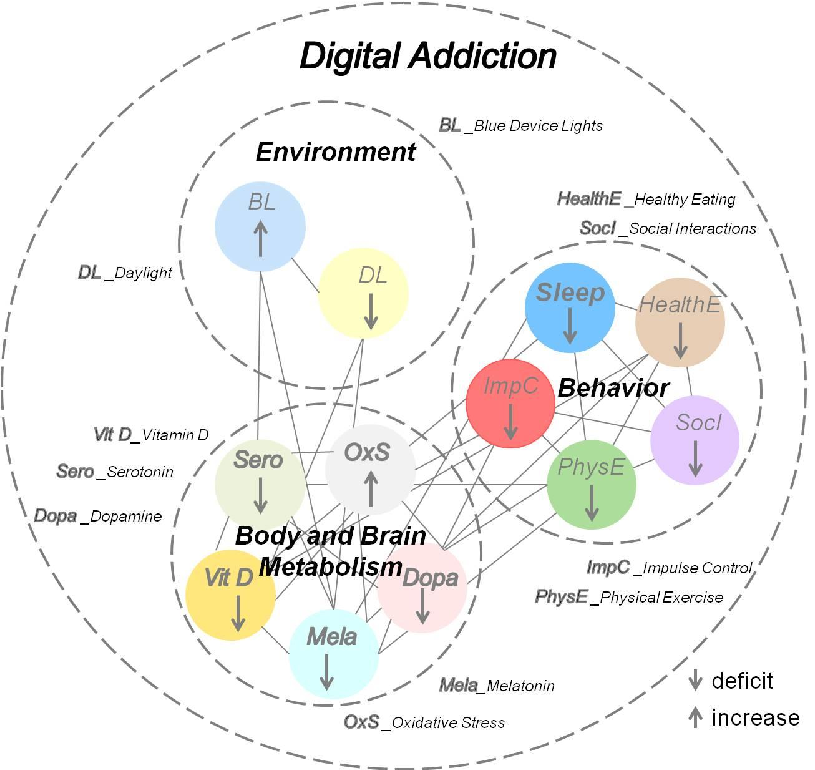
Neurobiological mechanisms linking excessive digital device use to disrupted sleep architecture are outlined, connecting screen-mediated blue light exposure and dopaminergic reward activation to circadian rhythm disturbances.
Digital Addiction and Sleep.
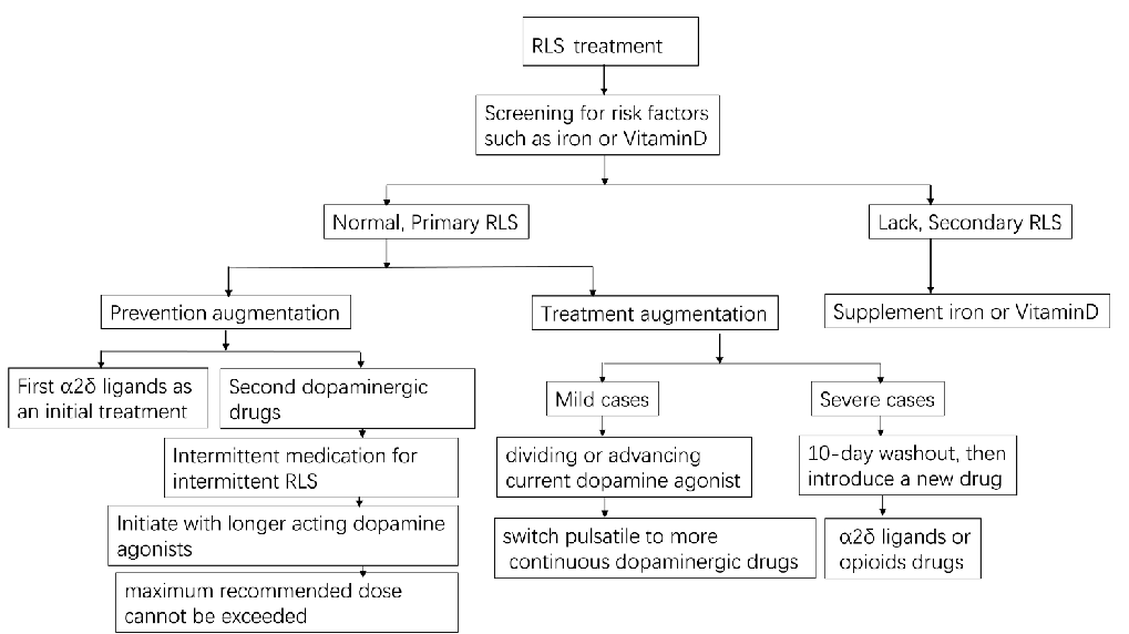
In mild RLS cases, dopamine agonist therapy management after augmentation is illustrated, showing dose adjustment strategies and transition to alternative medications including alpha-2-delta ligands and opioids for resistant cases.
Pharmacologic Treatment of Restless Legs Syndrome.
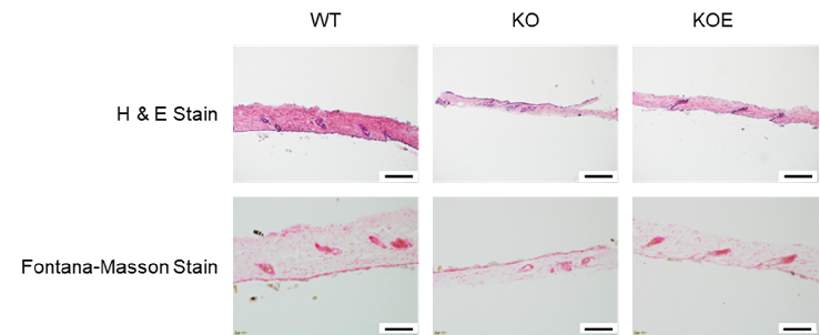
Molecular pathway analysis related to eggshell membrane effects on skin aging, depicting signaling cascades involved in keratinocyte differentiation or senescence.
Effects of Eggshell Membrane on Keratinocyte Differentiation and Skin Aging In Vitro …