Magnesium Glycinate Şekiller
10 hakemli araştırmalardan görseller
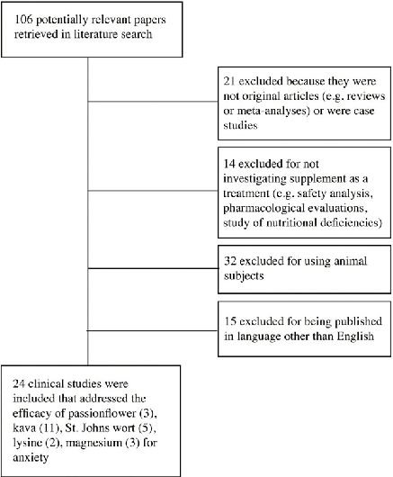
A PRISMA flow diagram outlines the systematic search and screening process for studies evaluating nutritional and herbal supplements for anxiety disorders. Database searches, inclusion criteria application, and final study selection are detailed.
Nutritional and herbal supplements for anxiety and anxiety-related disorders: systematic review.
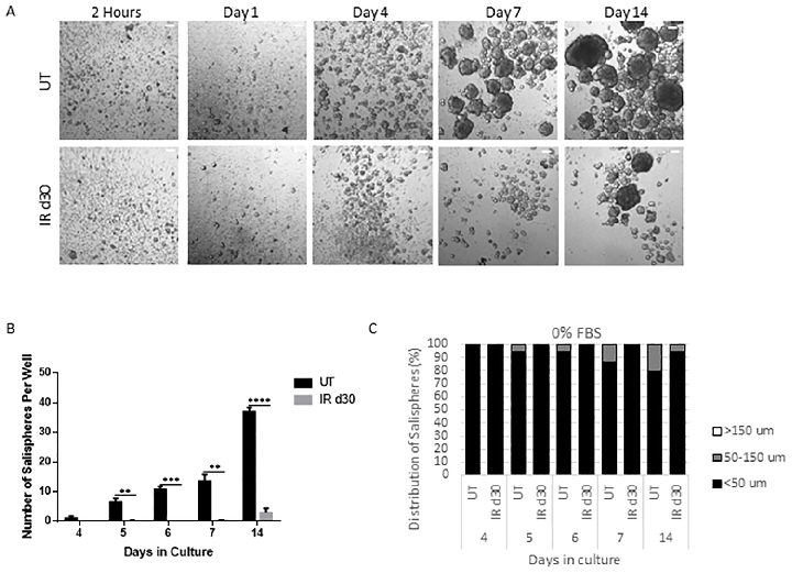
Sphere-forming efficiency of parotid-derived cells decreases significantly following a single 5 Gy radiation dose, quantified at day 30 post-irradiation.
Administration of growth factors promotes salisphere formation from irradiated parotid salivary glands.
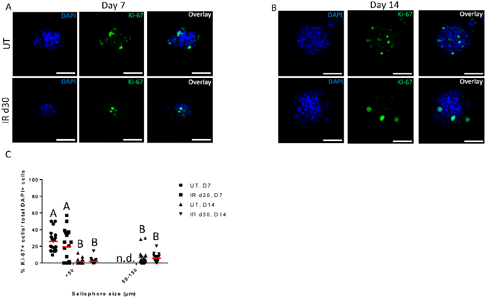
Proliferation rates of salisphere cultures from untreated and irradiated parotid glands are compared under serum-free conditions, showing similar growth kinetics despite radiation damage.
Administration of growth factors promotes salisphere formation from irradiated parotid salivary glands.
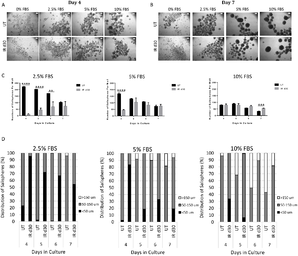
Fetal bovine serum supplementation increases the sphere-forming efficiency of irradiated parotid-derived cells, as shown by representative bright-field microscopy images and quantification.
Administration of growth factors promotes salisphere formation from irradiated parotid salivary glands.
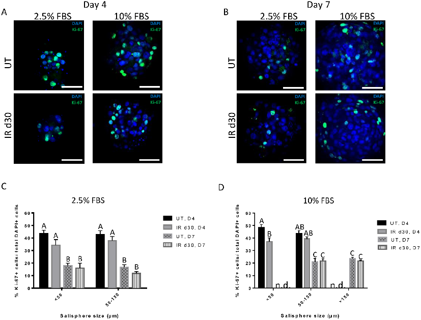
Proliferation assays confirm comparable growth rates between untreated and irradiated salisphere cultures supplemented with FBS, indicating that reduced sphere formation is not due to proliferation deficits.
Administration of growth factors promotes salisphere formation from irradiated parotid salivary glands.
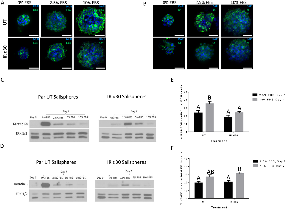
Expression of salivary stem and progenitor cell markers in salisphere cultures demonstrates that both untreated and irradiated cells maintain their progenitor phenotype.
Administration of growth factors promotes salisphere formation from irradiated parotid salivary glands.
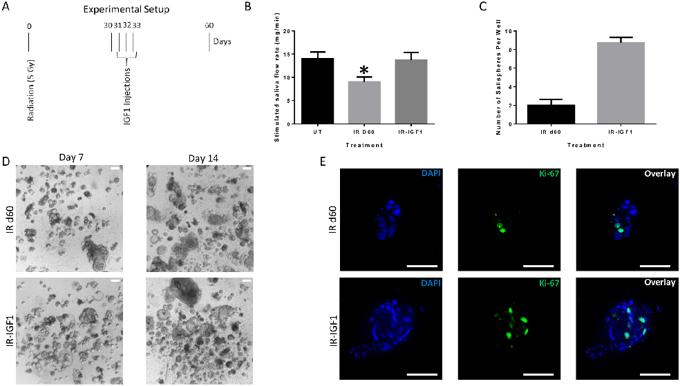
Post-radiation IGF-1 treatment significantly enhances sphere-forming efficiency of irradiated parotid cells, suggesting growth factor administration as a strategy to restore salivary function.
Administration of growth factors promotes salisphere formation from irradiated parotid salivary glands.
Skin hydration measurements in human subjects receiving oral eggshell membrane supplementation are tracked over the study period.
Effects of Eggshell Membrane on Keratinocyte Differentiation and Skin Aging In Vitro …
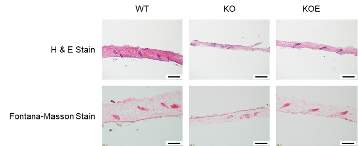
Histological sections of reconstructed skin models show improved epidermal architecture in eggshell membrane-treated constructs.
Effects of Eggshell Membrane on Keratinocyte Differentiation and Skin Aging In Vitro …
Molecular pathway analysis related to eggshell membrane effects on skin aging, depicting signaling cascades involved in keratinocyte differentiation or senescence.
Effects of Eggshell Membrane on Keratinocyte Differentiation and Skin Aging In Vitro …