Melatonin Şekiller
49 hakemli araştırmalardan görseller
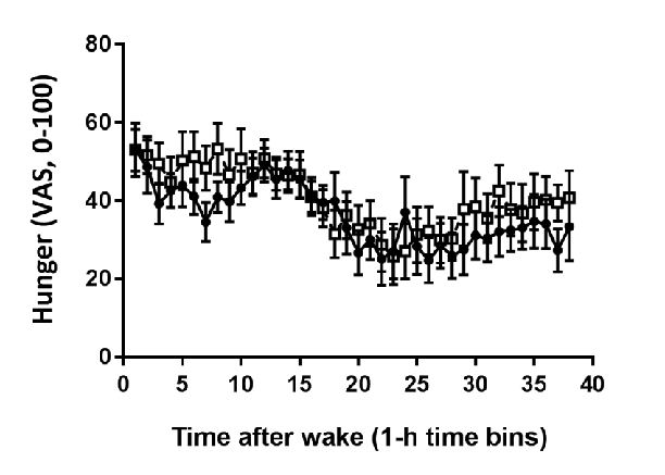
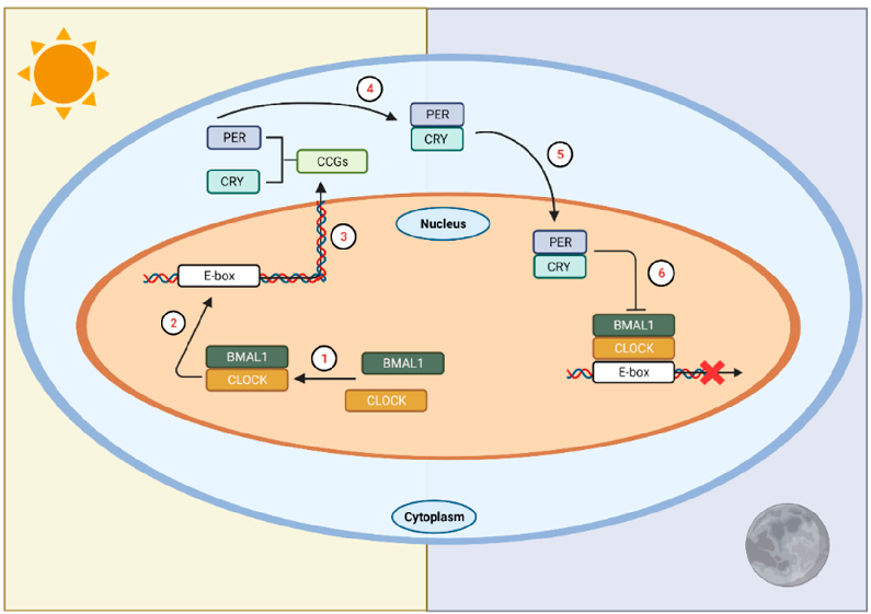
Circadian rhythm regulation involves a complex interplay between the central clock in the suprachiasmatic nuclei and peripheral oscillators throughout the body. Melatonin serves as the primary hormonal signal conveying darkness information to these systems.
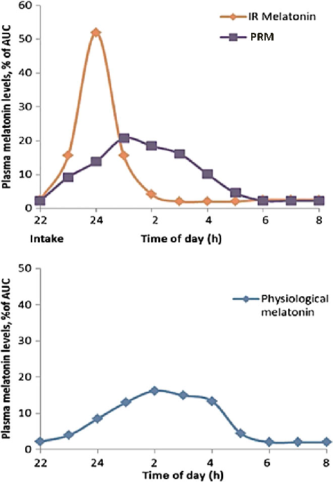
New perspectives on the role of melatonin in human sleep, circadian rhythms …
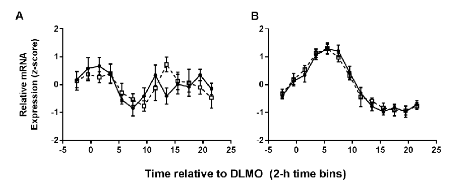
Exogenous melatonin administration influences circadian phase positioning, with the direction and magnitude of phase shifts depending on the timing of administration. Evening doses advance the circadian clock, while morning doses may cause phase delays.
New perspectives on the role of melatonin in human sleep, circadian rhythms …
Clinical applications of melatonin extend beyond sleep induction to include circadian rhythm resynchronization in jet lag, shift work disorder, and delayed sleep-wake phase disorder. Dose-response relationships vary across these conditions.
New perspectives on the role of melatonin in human sleep, circadian rhythms …
Hormonal response profiles to standardized meals are compared between normal and delayed meal timing conditions, revealing shifts in metabolic hormone rhythms.
Meal Timing Regulates the Human Circadian System.
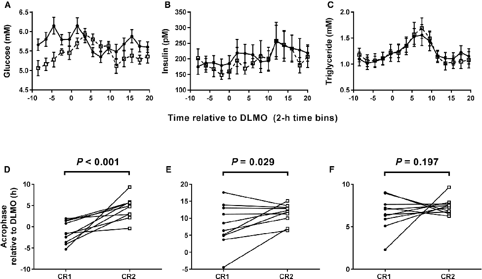
Average plasma glucose concentration under constant routine conditions is reduced following a 5-hour delay in meal times, suggesting meal timing significantly influences glucose metabolism rhythms.
Meal Timing Regulates the Human Circadian System.
Core body temperature rhythms are displayed across experimental conditions, demonstrating that meal timing shifts selectively affect peripheral but not central circadian markers.
Meal Timing Regulates the Human Circadian System.
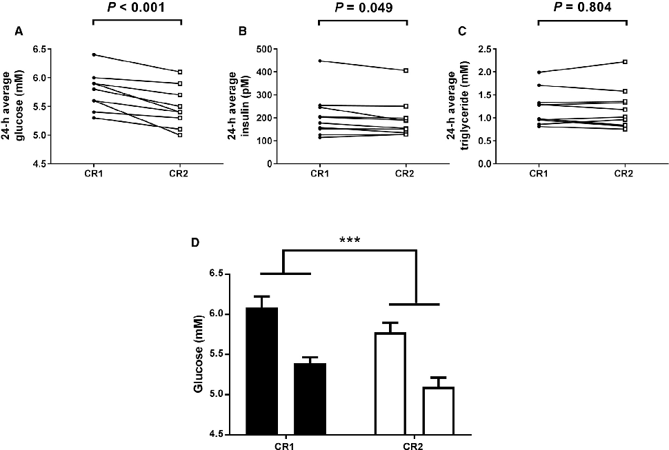
Plasma glucose concentrations during constant routine conditions show a significant reduction after participants experienced delayed meal times, indicating that meal scheduling influences circadian glucose regulation.
Meal Timing Regulates the Human Circadian System.
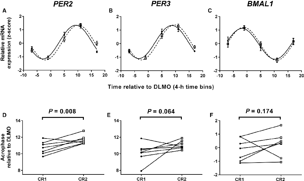
Clock gene expression patterns in adipose tissue are compared between the two meal timing conditions, revealing tissue-specific circadian phase shifts.
Meal Timing Regulates the Human Circadian System.
Per2 and other clock gene transcript levels measured from peripheral blood samples demonstrate delayed phase alignment consistent with the shifted meal schedule.
Meal Timing Regulates the Human Circadian System.
Summary model of how meal timing acts as a zeitgeber for peripheral clocks independent of the central suprachiasmatic nucleus pacemaker.
Meal Timing Regulates the Human Circadian System.
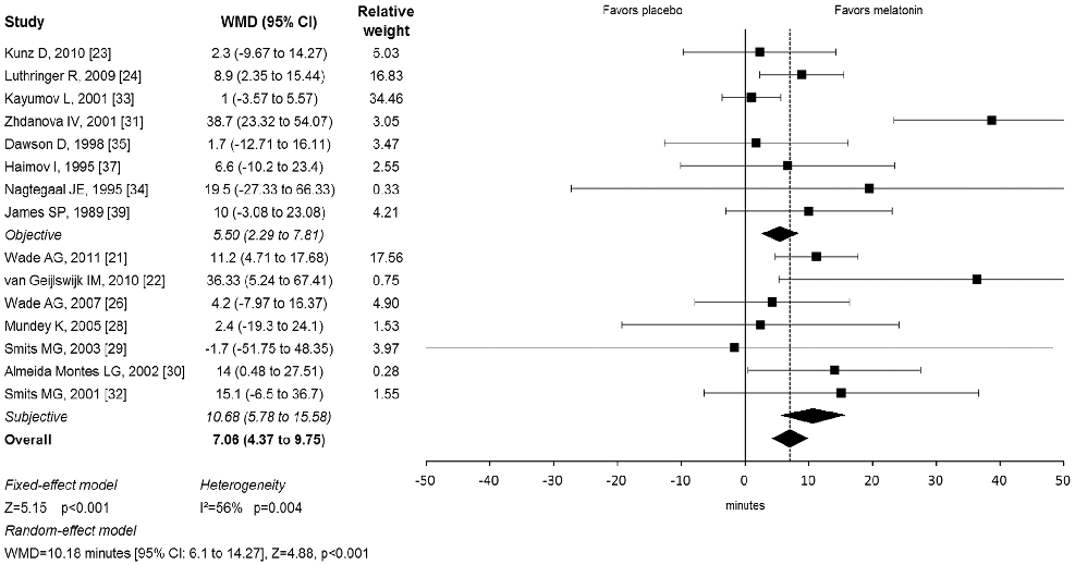
Forest plot from a meta-analysis of melatonin supplementation trials displays weighted mean differences in sleep onset latency, suggesting melatonin is associated with modest but statistically significant improvements in time to fall asleep.
Meta-analysis: melatonin for the treatment of primary sleep disorders.
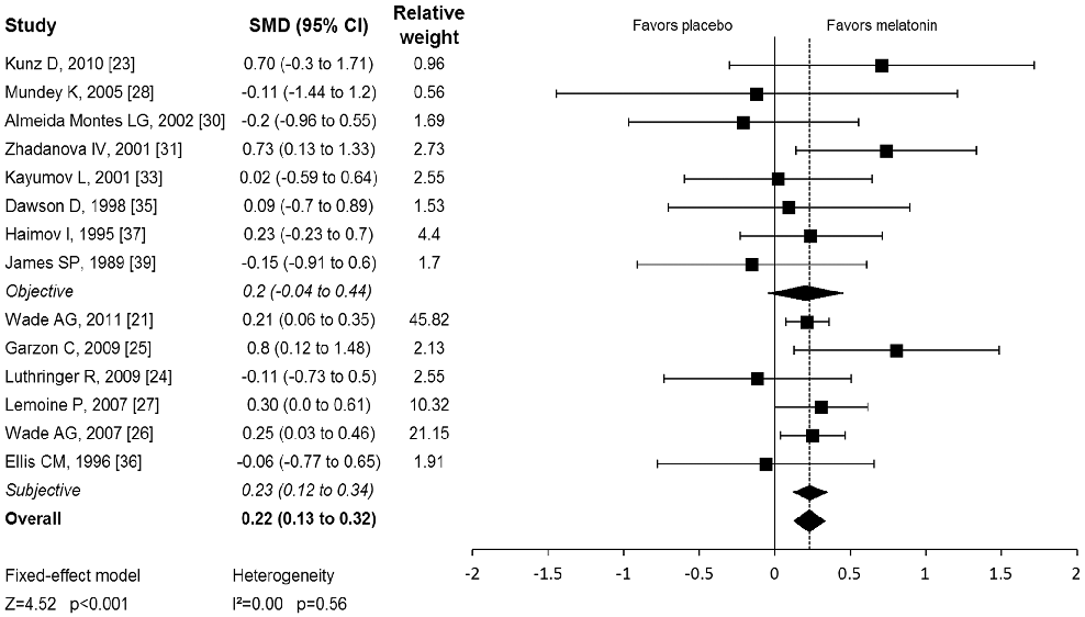
Pooled effect estimates for melatonin's impact on total sleep time and sleep quality across randomized controlled trials are presented, with subgroup analyses by dosage and duration.
Meta-analysis: melatonin for the treatment of primary sleep disorders.
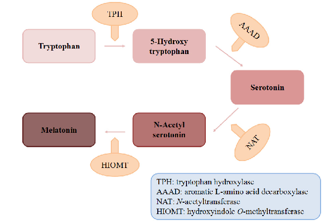
Biosynthetic pathway of melatonin from tryptophan is displayed, showing the sequential enzymatic steps through serotonin N-acetyltransferase and hydroxyindole-O-methyltransferase.
Dietary Sources and Bioactivities of Melatonin.
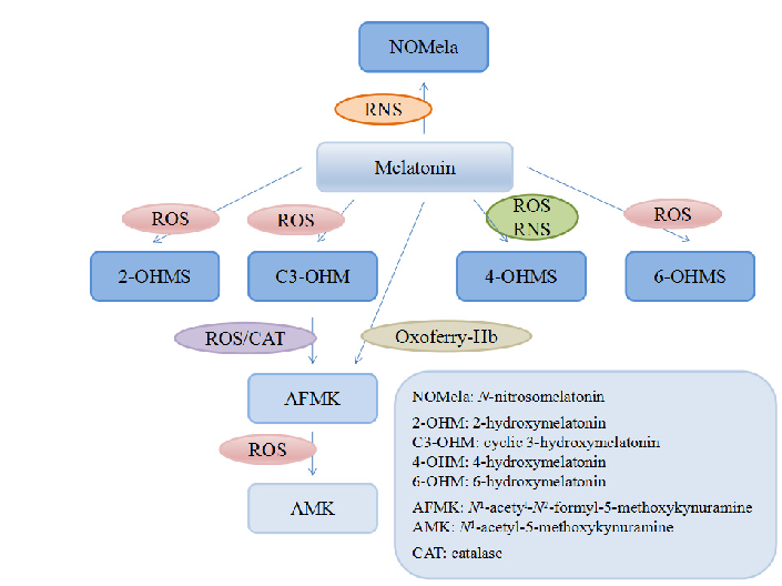
Melatonin and its metabolites including 6-hydroxymelatonin, AFMK, and AMK are structurally depicted, illustrating the biotransformation cascade.
Dietary Sources and Bioactivities of Melatonin.
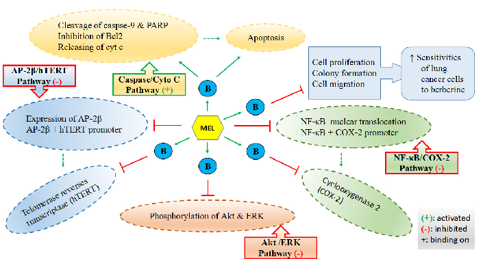
Mechanisms by which melatonin enhances lung cancer cell sensitivity to berberine are diagrammed, showing synergistic effects on apoptotic and autophagic pathways.
Dietary Sources and Bioactivities of Melatonin.
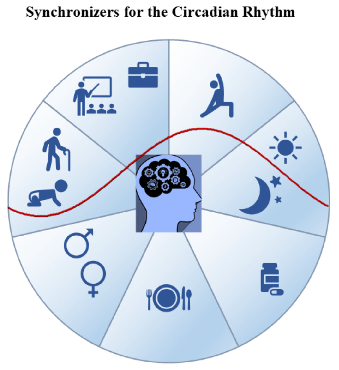
Factors influencing the rest-activity circadian rhythm and the sleep-wake cycle are mapped, encompassing light, melatonin, physical activity, and social timing cues.
Biological Rhythm and Chronotype: New Perspectives in Health.
PRISMA-style flow chart shows the systematic selection process for studies included in this review on biological rhythm and chronotype in health.
Biological Rhythm and Chronotype: New Perspectives in Health.
Metabolic consequences of circadian disruption include altered glucose tolerance, lipid metabolism, and energy balance. This figure summarizes the pathophysiological links between circadian misalignment and metabolic disease risk.
Circadian Rhythms Disrupted by Light at Night and Mistimed Food Intake Alter …
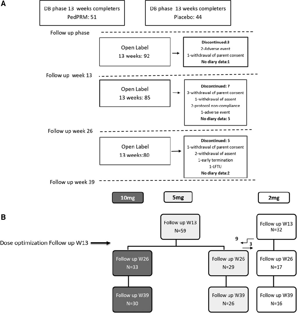
Study enrollment and participant flow for a long-term follow-up trial of prolonged-release melatonin (PedPRM) in children and adolescents with autism spectrum disorder and insomnia. The diagram tracks participants through screening, treatment phases, and follow-up periods.
Long-Term Efficacy and Safety of Pediatric Prolonged-Release Melatonin for Insomnia in Children …
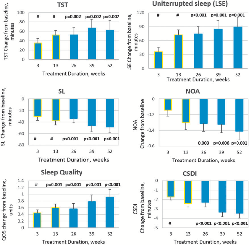
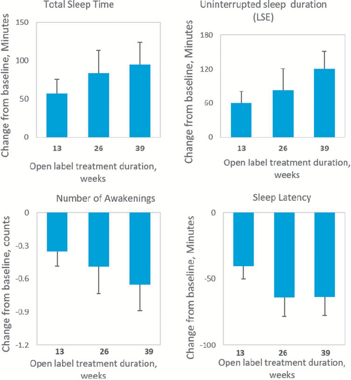
Efficacy outcomes showing changes in sleep parameters over the extended treatment period with pediatric prolonged-release melatonin in children with ASD. Long-term data indicate sustained improvements in sleep onset latency and total sleep time.
Long-Term Efficacy and Safety of Pediatric Prolonged-Release Melatonin for Insomnia in Children …
Safety assessment data from the open-label extension of PedPRM treatment in pediatric patients with autism spectrum disorder. Adverse event profiles suggest the prolonged-release melatonin formulation maintained an acceptable safety profile over the extended study period.
Long-Term Efficacy and Safety of Pediatric Prolonged-Release Melatonin for Insomnia in Children …
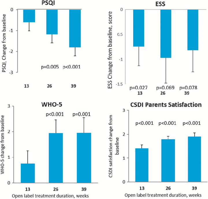
Summary of sleep quality measures or caregiver-reported outcomes during long-term PedPRM administration in children with ASD and comorbid insomnia. Results suggest continued therapeutic benefit with nightly melatonin use.
Long-Term Efficacy and Safety of Pediatric Prolonged-Release Melatonin for Insomnia in Children …
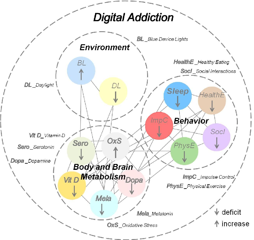
![Figure 1. The iRISA syndrome [76] in addiction is centrally controlled by dopamine in the brain, while asynchronization, presumed to be linked to cue sensitivity in digital addiction [50–54], is centrally controlled by serotonin. A deficit in both neurotra](https://pdfs.citedhealth.com/figures/35682491/101.png)
The impaired Response Inhibition and Salience Attribution (iRISA) syndrome model is illustrated, showing how dopamine-mediated reward pathways in the brain drive cue sensitivity in digital addiction and contribute to sleep disruption.
Digital Addiction and Sleep.
Neurobiological mechanisms linking excessive digital device use to disrupted sleep architecture are outlined, connecting screen-mediated blue light exposure and dopaminergic reward activation to circadian rhythm disturbances.
Digital Addiction and Sleep.
Sayfa 1 / 3